A “Glossary of nervous diseases” is not a single, universally defined title of a specific medical textbook or official document. Instead, it is a descriptive term used for reference materials that provide definitions and explanations of neurological disorders.
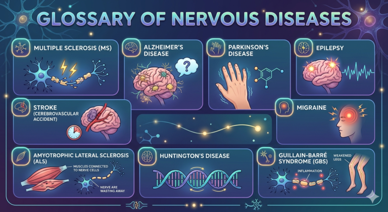
AMYOTROPHIC LATERAL SCLEROSIS is an organic disease of the brain and spinal cord, which is based on isolated damage to the motor neurons (nerve cells and their axons) innervating the muscles of the limbs, tongue, pharynx, and larynx. It usually begins at the age of 40-50 years. Characteristics are flaccid atrophic paresis and paralysis of the arms, combined with increased tendon reflexes, spastic paresis of the legs, speech disorders (dysarthria), and swallowing disorders.
The course of the disease is progressive; the disease ends fatally in 2-5 years. The causes of the disease are unknown. Since there is no specific treatment, proserin, oxazyl (kalimin), vitamins E, B1, and B12 are prescribed in courses. Interferon and interferonogens are used. Physiotherapy, massage, and therapeutic exercises are widely used. In the development of widespread atrophy, retabolil and nerobol are prescribed.
APRAXIA is the inability to perform complex, purposeful actions (the patient forgot how to dress, button up, make the bed). It is observed with focal brain lesions, trauma, and cerebrovascular disorders.
ARACHNOIDITIS is an inflammation of the arachnoid mater of the brain or spinal cord, in which adhesions occur between the arachnoid and choroidal mater, followed by circulatory impairment and the formation of cystic cavities. It can occur with general infections, otitis, inflammatory diseases of the paranasal sinuses, brain tumors, and traumatic brain injury.
Symptoms: Local and diffuse headaches, nausea, sometimes mild meningeal syndrome, and subfebrile temperature. When arachnoiditis is predominantly localized at the base of the brain and the optic nerves are involved, visual acuity and visual field are reduced. Arachnoiditis in other locations presents with a clinical picture resembling a tumor in the corresponding location, and a correct diagnosis is possible only with radiography, tomography, and cerebrospinal fluid analysis.
Treatment. In the acute phase, treatment is carried out in a hospital. Corticosteroids, veroshpiron, and B vitamins are prescribed. Neurosurgical treatment (adhesion dissection) is performed as indicated.
ATAXIA is a disorder of motor coordination due to damage to the vestibular system, cerebellum, etc. It manifests as impaired balance when standing (static ataxia) and impaired motor coordination (dynamic ataxia). If ataxia is suspected, ask the patient to stand with their arms extended forward, eyes closed, and legs together. Then, touch the tip of their nose with their finger, or, lying on their back, touch the knee of the opposite leg with their heel. If the person is unable to perform these tasks, consult a doctor.
ATHETOSIS is a type of hyperkinesia characterized by slow, involuntary, forced movements due to tonic muscle contractions. Athetoid movements may occur not only in limited muscle groups (face, arm, leg), but also involve the entire musculature. Limited athetosis occurs with toxic brain damage (carbon monoxide, manganese, carbon disulfide), while generalized athetosis is characteristic of certain lesions of the subcortical regions of the brain (hepatocerebral dystrophy, etc.). Double athetosis is generalized slow, involuntary movements involving the muscles of the extremities, trunk, and face on both sides. Typically, it results from perinatal brain damage in children who have suffered fetal asphyxia.
APHASIA is a speech disorder caused by organic brain damage. Aphasia can be motor, sensory, or amnestic. With motor aphasia, patients lose the ability to speak but understand the speech of others. With sensory aphasia, patients lose the ability to understand speech but can pronounce individual words and phrases. With amnestic aphasia, patients know the purpose of objects but cannot name them correctly.
INSOMNIA is a very common ailment, characterized by difficulties with falling asleep or staying asleep, lasting at least 3 weeks. There are three main types of insomnia: sleep onset disorder, night waking, and early awakening.
1. “I go to bed and can’t fall back asleep for a very long time.”
This condition can be contributed to by:
– Stress. Emotional stress includes anxiety, depression, self-pity, anger, and guilt. Its causes are varied.
– Unresolved issues, mental imbalance, various physical disorders, and neurological problems.
– Diand gestive and nutritional problems. Consuming stimulating foods and drinks (strong tea, coffee) before bed, or having a too large and high-calorie dinner.
– Incorrect sleep schedule. Daytime naps (after lunch, in the evening) and, as a result, a lesser need for sleep at night.
– Big changes in life, which can be both good and bad, such as moving to a new place of residence, divorce, the death of loved ones, a change of job, etc.
– Disruption of the biological clock (night work and jet lag).
– External stimuli, such as noise.
2. “I often wake up in the middle of the night and lie there, unable to fall asleep.”
Interrupted sleep at night can be caused by all of the above reasons, as well as:
– Irritation and anger.
– Severe alcohol intoxication, or, conversely, abstinence from alcohol and medications – on the advice of a doctor or for another reason.
– Nightmares, fear of having a nightmare.
– A sedentary lifestyle, insufficient fatigue during the day.
3. “I wake up very early and cannot fall asleep again.”
– Severe depression.
– Alcoholism.
– Addiction to sleeping pills.
Treatment. It’s important to understand the underlying causes of insomnia and try to eliminate them. To avoid sleepless nights, try following these guidelines in the evening.
Never deal with troubling problems before bed or in bed. It’s best to focus on them during the day or early evening. Avoid stimulating drinks (tea, coffee, alcohol) in the evening. Dinner should be light, but avoid raw vegetables and fruits, as well as black bread, as these can cause stomach rumbles, which can also interfere with sleep. Avoid any stimulating activity in the evening (vigorous exercise, work, arguments, etc.). Spend the last hour preparing for bed, including relaxation exercises and a warm (not hot!) bath with pine extract. You can also drink an infusion of mint or motherwort. Create a cozy, restful environment in your bedroom. Read Pushkin before bed (the rhythm of poetry matches the natural rhythms of the heart and brain, which calms an overstimulated mind). Try to eliminate noise that can disrupt your sleep. It is often easier to fall asleep if a small lamp is on in the room.
Even if you’re a very busy person and used to constantly doing something or rushing somewhere, try to do nothing in the evening and set aside time and space to think, dream, or simply take a walk. This may be just what you need to fall asleep peacefully. If you can’t sleep on your own, you should consult a doctor. Don’t start taking sleeping pills without consulting a doctor, as they quickly become addictive, making it much more difficult to combat insomnia.
BULBAR PALSY is a symptom complex consisting of swallowing and speech difficulties. It occurs with damage to the medulla oblongata and the cranial nerve nuclei, pathways, and reticular formation located there. Swallowing difficulties, choking and coughing when eating, a, nd breathing difficulties are observed. Speech may be nasal, pronunciation may be impaired, and hiccups, vomiting, cardiovascular problems, and arrhythmias may occur. If bulbar syndrome develops, immediate hospitalization in a neurological hospital is necessary.
AUTONOMIC DYSTONIA is a symptom complex characterized by functional disorders in various parts of the autonomic nervous system. Autonomic dystonia is most often observed in neuroses and manifests itself through various disturbances of the cardiovascular, respiratory, and gastrointestinal tracts. Cardiovascular disturbances include palpitations, increased or decreased blood pressure, pallor, and sweating; digestive disturbances include loss of appetite, belching, difficulty swallowing, nausea, and hiccups; and respiratory disturbances include shortness of breath and chest tightness. Characteristic complaints include fatigue, restless sleep, and emotional instability. A slight and transient increase in blood pressure and tachycardia is also noted.
The manifestations of autonomic dystonia are very diverse and can resemble several different diseases at once. Patients’ behavior is often persistent, and the multitude of incongruous complaints they present can bewilder the physician. Sometimes, even with a diagnosis of autonomic dysfunction, the underlying condition remains undetected. Therefore, a diagnosis of autonomic dysfunction is one of exclusion and is made only after an ECG, EEG, visual field examination, examinations by an ophthalmologist, endocrinologist, and psychotherapist, as well as urine and blood tests, have been performed.
Treatment: A range of health and hygiene measures, measured physical activity, spa and physiotherapy, autogenic training, and other psychotherapeutic methods are recommended. Medications that normalize nervous processes are prescribed, including valerian, bromine preparations, and tranquilizers.
HYDROCEPHALY (water on the brain) is the accumulation of excess cerebrospinal fluid in the cranial cavity. Hydrocephalus can be congenital or acquired. Congenital hydrocephalus develops as a result of intrauterine infections and intoxications. Children with congenital hydrocephalus have dilated cranial sutures, a tense, non-pulsating fontanelle, and mental retardation, strabismus, and other symptoms. Acquired hydrocephalus develops after infections and head injuries. It is characterized by increased intracranial pressure and presents with headache and vomiting.
Treatment: It is necessary to determine the cause of hydrocephalus and treat the underlying condition. Diuretics, mannitol, glycerin, and glucocorticoid hormones are used to reduce intracranial pressure. In some cases, surgical treatment is indicated.
HEADACHE: A common symptom of various organic and functional nervous system diseases. Headaches of varying intensity and location are observed with damage to the meninges, increased intracranial pressure, trigeminal neuralgia, head injuries, inflammation of the paranasal sinuses, hypertension, atherosclerosis, and eye diseases. Headaches often occur with diseases of the internal organs (liver and kidney disease, anemia, chronic poisoning with industrial toxins). Headaches can be associated with fatigue, emotional stress, etc.
Treatment. It is necessary to determine and treat the underlying disease. In addition, painkillers (citramon, fentanyl, analgin, acetylsalicylic acid, etc.) are prescribed.
DIZZINESS is a sensation of apparent movement of the body in space or of surrounding objects. It is often a symptom of damage to the vestibular system, brainstem, cerebellum, or cerebral cortex. It may be accompanied by nystagmus, vomiting, gait disturbance, and pale skin. Increased blood pressure accompanying dizziness often indicates vascular disorders. Dizziness as a symptom of a disease may be caused by an inner ear disorder, when an inflammatory or tuorous process affects the vestibular system. Such patients typically suffer from an inner ear infection (otitis media) for a long time; they initially experience hearing loss, followed by dizziness. With a tumor, attacks occur alongside hearing loss and ringing in the ear, and the dizziness gradually increases in frequency and intensity.
Dizziness is often experienced with a sharp turn of the head to the side, when rising abruptly in the morning, when tilting the head back (while wallpapering, hanging laundry), or when tilting the head to the side or forward for a long time (weeding, car repair, sleeping long hours on a commuter train, etc.). These dizziness symptoms are caused by a spasm or compression of the vertebral artery, which supplies blood to the areas of the brain responsible for balance. Dizziness can occur with high or low blood pressure, poisoning, fluid loss (vomiting, diarrhea), or blood loss (bleeding). Dizziness is accompanied by dizziness and anemia (a lack of red blood cells) and a high fever.
Dizziness can also occur in a perfectly healthy person, for example, if they spin around for a long time, look down from a great height, or look at the clouds.
First aid. If the patient experiences dizziness, they should be placed on their back with their head, neck, and shoulders supported by a pillow. This prevents kinking of the vertebral arteries. Avoid turning the head to the side. Open windows, ventilate the room, and apply a cold compress to the forehead, perhaps slightly soaked in vinegar. Measure the patient’s blood pressure and temperature, and take steps to normalize them. If the heart rate exceeds 100 beats per minute or irregular heartbeats develop, or if dizziness is accompanied by nausea and repeated (more than three) vomiting, call an ambulance. The cause of the dizziness must be determined and the underlying condition treated.
CEREBRAL PALSY is a group of motor dysfunction syndromes that result from brain damage in the womb, during birth, or early in life. It most often occurs as a result of infections, intoxications, and birth injuries. Children with cerebral palsy experience poor physical development and are late in learning to sit, walk, and talk. The primary impairments manifest as paresis, paralysis, and hyperkinesis. Mental disorders are rare.
Treatment is comprehensive and aimed at restoring movement and speech disorders. Massage, therapeutic exercise, and speech therapy are regularly performed. Medications that normalize muscle tone (elenium, baclofen, etc.) and metabolic drugs (cerebrolysin, piracetam, etc.) are prescribed. With age, with consistent treatment, motor defects may diminish and, in some cases, disappear completely.
STUTTERING (LOGONEUROSIS) is a disorder characterized by no pathological changes in the articulatory (speech-conducting) apparatus (larynx, vocal cords, lungs, lips, teeth, tongue) and no damage to the area of the brain that controls speech. However, the speech control system functions incoherently with the articulatory apparatus. Stuttering is the result of a spasm in one or more organs involved in speech production. The flow of words is suddenly interrupted, resulting in a pause. Sometimes this occurs as a result of the rapid repetition of a sound after the initial pause. There are many degrees of stuttering. It can range from a slight inability to pronounce final letters or syllables to a situation where a spasm completely paralyzes the muscles of the tongue and larynx. Stuttering is often caused by emotional stress.
Stutterers typically experience the most trouble with explosive consonants: “b,” “p,” “d,” “t,” “k,” and the hard “g.” These consonants are formed by stopping the flow of air and pressing on the lips, followed by a sudden burst of airflow and its release through the open lips. Stuttering is often corrected by following reading rules and slowly and carefully pronouncing each syllable.
Stuttering typically occurs at a young age, when speech automatisms are not fully developed. Stressful situations experienced during childhood (fright, the death of a loved one or a beloved pet, parental divorce, constant arguments, etc.) contribute to the development of stuttering. True stuttering intensifies with anxiety and subsides in a calm environment. People who stutter are usually good singers and do not stutter while singing.
Treatment. Speech therapists and psychologists treat stuttering, as the disease is primarily functional in nature and is not associated with true damage to the speech apparatus. The prognosis is usually favorable.
A STROKE is an acute cerebrovascular accident that most often develops as a complication of hypertension and cerebral atherosclerosis. A distinction is made between hemorrhagic (intracerebral hemorrhage, subarachnoid hemorrhage) and ischemic stroke.
HEMORRHAGIC STROKE (CEREBRAL HEMORRHAGE)A stroke occurs as a result of an artery rupture due to fluctuations in blood pressure or changes in the functional state of the vessels. Emotional stress or physical exertion can trigger the development of a stroke. Characterized by sudden onset, facial flushing, severe headache, loss of consciousness, repeated vomiting, and paralysis of the limbs on one side. On the side of paralysis, the corner of the mouth is drooping; sometimes, the head and eyes turn toward the side of paralysis. Blood pressure reaches 180/100 mmHg and above, and there is tachycardia. After a few hours, body temperature rises to 37-39 °C, and leukocytosis appears in the blood. In cases of extensive hemorrhage, accompanied by progressive respiratory and cardiac impairment, the patient may die.
Treatment: In most cases, patients are hospitalized. First, the airways are cleared of mucus and vomit. The head should be elevated. To compensate for cardiac activity, the administration of cardiac glycosides is indicated. In the presence of very high blood pressure, it is necessary to reduce it under constant medical supervision to the patient’s usual values (usually not lower than 150-160/90 mmHg). Lasix, dibazol, clonidine, papaverine, and no-shpa are administered intramuscularly. Slow intravenous administration of euphyllin is effective in the first hours of the disease. In severe cases, glucocorticoid hormones (hydrocortisone, dexamethasone) are used. It is necessary to monitor bladder and bowel function.
SUBARACHNOID HEMORRHAGE . Occurs with a ruptured aneurysm of the cerebral vessels, less often with hypertension and other vascular disorders. Characteristic symptoms include a sudden, severe headache “like a blow to the back of the head”, vomiting, epileptiform seizures, and motor restlessness. Clouding of consciousness and a stupor are observed. Meningeal syndrome manifests itself.
Treatment: The patient must be hospitalized. Strict bed rest is required for 4 weeks. Epsilon-aminocaproic acid and sterile glycerin solution are administered intravenously for the first 7-10 days. Analgesics (baralgin, analgin) are prescribed. During the first 4 weeks, therapeutic exercises and massage are performed in bed, then, depending on the patient’s condition, sitting and walking are allowed. Bladder and bowel function must be regulated. For the first 8 weeks, the patient should avoid any physical exertion or straining to prevent rebleeding. If an aneurysm ruptures, surgical treatment is performed.
ISCHEMIC STROKE (CEREBRAL INFARCTION)It most often develops in old age as a result of blockage (thrombus, embolus) or severe narrowing of atherosclerotic blood vessels in the brain. Emotional stress, fatigue, and infectious diseases can trigger the condition. The disease usually develops gradually, with consciousness preserved. Symptoms include headaches, dizziness, general weakness, pale skin, and transient numbness in the arm or leg. The malaise progresses, and over the course of several minutes, an hour, or sometimes a day, the arm and leg become increasingly heavy, then weaken sharply and become completely immobile. If the ischemic process develops at night, the patient may feel unable to stand in the morning, as the arm and leg are immobile. If paralysis affects the right limbs, speech may be lost or severely impaired. If the left limbs are affected, speech is usually not impaired. Cerebral infarction is also characterized by visual impairment and difficulty swallowing.
Treatment. Slow intravenous administration of aminophylline solution with corglycon and glucose solution, as well as intravenous drip infusion of rheopolyglucin, are recommended. Dexamethasone is used as indicated. Antiplatelet agents (acetylsalicylic acid or Trental) are prescribed starting on the 4th to 5th day of illness. The patient should undergo massage and therapeutic exercises, monitor bladder and bowel function, and prevent bedsores by regularly turning the patient. If the disease is mild and cardiac function is satisfactory, the patient is allowed to stand and walk starting on the 5th to 7th day of illness.
COMA is a state of unconsciousness caused by dysfunction of the brainstem. It is characterized by respiratory and cardiac dysfunction, lack of response to various external stimuli, and lack of active movement. It can occur with traumatic brain injury, brain tumors, diabetes, stroke, uremia, and alcohol and drug intoxication. In mild coma, patients respond to pain stimuli, and pupillary and corneal reflexes are preserved. In deep coma, areflexia, atony, mydriasis, and severe respiratory and circulatory impairment are observed. In the terminal stage of coma, the patient’s vital functions are maintained only through artificial ventilation and cardiac stimulation. A comatose patient requires immediate hospitalization.
LUMBAGO (LUMBAGO) is a pain that occurs acutely in the lumbar region during physical exertion (sudden movement, lifting something heavy).
Symptoms The disease develops acutely: during exertion, severe pain in the lower back suddenly appears, the patient is unable to bend or straighten, freezes in one position, and sometimes falls due to the pain. The pain increases sharply with any movement, coughing, or sneezing. Palpation reveals tension in the back muscles. After a few days, the pain subsides, and the range of motion in the spine is gradually restored.
Treatment: Bed rest and analgesics (analgin, baralgin, fentanyl) are prescribed. Rubbing with Voltaren ointment, Menovazine, and mustard plasters is indicated.
MENINGEAL SYNDROME. Meningeal irritation syndrome may be observed with intoxication, general infections, increased intracranial pressure, subarachnoid hemorrhage, and meningitis. Irritation of the nerve roots causes reflex muscle tension (stiff neck – limitation or inability to bend the head forward, Kernig’s sign – inability to straighten the leg at the knee when bending the leg at a right angle at the hip). Important components of meningeal syndrome include headache, sometimes vomiting, increased sensitivity to light and noise, and possible mental disorders.
MENINGITIS is an inflammation of the pia mater of the brain and spinal cord. A distinction is made between primary and secondary meningitis. Primary (epidemic cerebrospinal) meningitis is caused by meningococcus. Secondary meningitis often develops as a complication of common infectious diseases (otitis, furuncles, influenza, mumps) and is caused by various pathogens (coccal flora, viruses, etc.).
Symptoms. The clinical picture of both forms of meningitis is similar. The onset of the disease is acute, body temperature rises to 39-40 °C, severe headache, vomiting, agitation, and often confusion and delirium occur. Meningeal syndrome (see) of varying severity is observed. Due to the involvement of the cranial nerves, strabismus, impaired convergence and accommodation, nystagmus, and anisocoria may occur. The blood shows neutrophilic leukocytosis, and ESR is accelerated. The cerebrospinal fluid is turbid, flows in a stream, contains many proteins, a large number of neutrophilic leukocytes, and meningococci. Possible complications: infectious-toxic shock, cerebral edema.
Treatment. Patients are hospitalized. Etiotropic, detoxifying, and dehydrating therapy are carried out. Benzylpenicillin is prescribed intramuscularly 6-8 times a day in high doses for 7-10 days. In case of penicillin intolerance or pathogen resistance to it, ampicillin, chloramphenicol, oxacillin, tetracycline, and metacycline are prescribed. And for detoxification, a 5% glucose solution, isotonic sodium chloride solution, plasma, albumin, etc., are administered intravenously. To prevent cerebral edema, Lasix, mannitol, etc., are administered. In severe cases, corticosteroids are indicated. For the treatment of convulsive syndrome, seduxen (diazepam) and sodium oxybutyrate are used. Patients are discharged from the hospital after receiving negative results of two bacteriological tests.
MIGRAINE– a disease characterized by attacks of headache, predominantly on one side of the head (hemicrania). Migraines are more common in young women. An attack is preceded by 10-20 minutes of malaise, irritability, heaviness in the head, drowsiness, and flashing spots before the eyes. The pain is throbbing, accompanied by redness or paleness of the face, redness of the eyeballs, nausea, and sometimes vomiting. An attack lasts from several hours to several days. The disease is not progressive, is not accompanied by signs of organic brain damage, and attacks become less intense with age.
Treatment: Since there is no consensus on the true nature of the disease, symptomatic treatments are used. Some patients respond to heat (heating pads on the feet, mustard plasters), others to cold, others to tightly wrapping the head with a towel, etc. During an attack, the patient requires absolute rest. Prescribed medications include sedatives (valerian, bromine preparations), tranquilizers (meprotan, elenium), antispasmodics and vasodilators (dibazol, papaverine), and analgesics (phenacetin, amidopyrine, novomigrofen). Diphenin, phenobarbital, and anaprilin reduce attacks. General health measures are recommended (spending time outdoors, adequate sleep, a work-rest schedule, and exercise).
MYELITIS is an inflammatory disease of the spinal cord. It can occur as a complication of general infectious diseases or traumatic injuries to the optic or spinal cord. Focal myelitis is characterized by acute onset, fever, back and chest pain, numbness and weakness in the legs, and urinary retention. Severe forms can cause limb paralysis, conductive sensory disturbances, and pelvic dysfunction.
Treatment is aimed at the underlying condition, of which myelitis is a complication. In most cases, patients require hospitalization. Antibiotics, analgesics, sedatives, anti-inflammatory drugs, and vitamins are used. Surgery is performed if necessary.
NEURALGIA is a peripheral nerve disorder most often caused by cold, infection, intoxication, trauma, or metabolic disorders. Neuralgia is characterized by paroxysmal pain of a stabbing or shooting nature, radiating widely along the sensory nerve’s innervation zone.
Trigeminal neuralgia most often occurs with various inflammatory diseases of the teeth, jaws, and paranasal sinuses. It is characterized by intense attacks of spontaneous facial pain on one side, lasting from a few seconds to 1-2 minutes. The attack occurs spontaneously and is often accompanied by facial muscle spasms. Examination sometimes reveals tenderness at the trigeminal nerve exit points, with “trigger” zones, where light touch triggers an attack.
Occipital neuralgia is characterized by pain in the back of the head. They are pounding and aching in nature, gradually spreading forward across the scalp, radiating from the back of the head to the temple and eye. The pain intensifies with head movements, palpation of the neck, sneezing, and coughing, and often causes nausea and vomiting.
Intercostal neuralgia is accompanied by pain along the intercostal space, usually one, but the second and third intercostal spaces can be affected. The pain is constant and aching. It intensifies with walking, breathing, turning the torso, and palpation of the ribs and intercostal spaces.
Treatment depends on the nature of the neuralgia and is supervised by a physician. Anticonvulsants, sedatives (seduxen, trioxazine), vitamins B1, B12, niacin, and physical therapy (UHF, diadynamic currents, electrophoresis with novocaine) are prescribed. Acupuncture is also effective. If conservative therapy is ineffective, surgical treatment is indicated.
NEURITIS is an inflammation of the nerve. (Neuritis is an outdated term; the term “neuropathy” is now commonly used, as most peripheral nerve lesions are non-inflammatory.) In clinical practice, neuropathies of the facial, radial, and peroneal nerves are most common.
FACIAL NERVE NEUROPATHY– most often of a vascular origin. It occurs after hypothermia, with peak incidence in September and March, i.e., when the off-season winds begin, and people go about without hats and scarves, wearing light raincoats, and are often exposed to drafts. The facial nerve exits the cranial cavity in the area behind the ear. When the head becomes too cold, a spasm of the blood vessels supplying the nerve occurs. Deterioration in nutrition leads to swelling and wedging of the nerve within the walls of the bony canal, which further impairs the nerve’s supply. The patient, upon waking in the morning, notes a slight aching pain behind the ear. When attempting to brush their teeth, they notice water leaking from the corner of their mouth. While eating breakfast, they notice food constantly getting between their cheek and teeth, interfering with chewing. Approaching a mirror, they notice that their face is immobilized on one side, and their mouth is pulled toward the opposite (healthy) side. The eye on the affected side does not close, the forehead does not wrinkle, and the corner of the mouth does not move when attempting to smile. Facial sensitivity (when pricking with a needle) remains.
Similar symptoms can be caused by conditions such as inflammation of the inner ear, a tumor of the auditory nerve, multiple sclerosis, a brain tumor, a stroke in the brainstem, and diabetes.
RADIAL NERVE NEUROPATHY. Typical complaints include the inability to straighten the fingers and raise the hand upward, although the hand can form a fist comfortably. The patient cannot clap when resting their hand on the table, nor can they tap the surface with their fingers. These disorders typically occur after heavy alcohol consumption the night before and an uncomfortable night’s sleep. For example, if the patient sleeps without a pillow, with their arm tucked under them, or if their spouse sleeps on their shoulder at night.
PERONEAL NERVE NEUROPATHY. Complaints include the inability to stand on the heels, the inability to put on slippers independently, and a “hanging” sensation in the foot. When walking, the patient stumbles on level ground, and their gait is also unusual: they try to lift or kick their leg forward and upward to avoid catching the floor. This disorder most often occurs after severe radiculitis, hypothermia, allergic reactions (such as bee stings), chemical poisoning (such as alcohol or insect repellents), atherosclerosis of the lower extremities, diabetes, and prolonged forced sitting, especially with their legs crossed.
Treatment: The sooner the patient seeks medical attention, the better the chances of success. If treatment for facial nerve neuropathy is not taken within the first 3-4 days, the face will remain immobile forever. Dry heat, aspirin, nicotinic acid, curantil, hormones, adhesive bandage traction, electrical stimulation, and acupuncture are used. If facial nerve neuropathy is a symptom of another condition, the underlying cause is treated first, but all nerve restoration measures are simultaneously implemented to ensure time is not lost.
Similar treatment is used for radial and peroneal nerve neuropathy.
FAINTING is a short-term drop in vascular tone, accompanied by cerebral anemia. It manifests as sudden weakness, dizziness, and loss of consciousness (for a few seconds or minutes). It is observed with certain cardiovascular diseases, blood loss, acute pain, severe anxiety, etc.
First aid: First, it is necessary to determine whether consciousness has been lost. To do this, the patient’s name is called loudly, their cheeks are slapped, and the pupils are dilated and reactive to light. This information will also be useful for the physician at the clinic or emergency room. The patient is placed with their legs elevated, loosened from any tight clothing, their face is splashed with cold water, and a cotton ball soaked in ammonia or cologne is given to them to smell. Simple fainting does not require treatment, but the patient may vomit during it, and a fall can break dentures. Fragments of dentures and food may enter the trachea and bronchi, which can lead to death by suffocation. The patient should be turned on their side, dentures removed, vomit removed, and given a cotton swab soaked in ammonia to sniff. If consciousness is not restored within 5-7 minutes, and especially if the patient has hit their head, immediate hospitalization is necessary.
BRAIN TUMORS can be primary (from the membranes or brain tissue) or secondary (metastatic). General cerebral symptoms include headache, nausea, vomiting, bradycardia, epileptic seizures, mental disturbances, and constant fatigue. Symptoms of focal lesions depend on the tumor’s location and may include paresis, paralysis, speech disorders, visual impairment, impaired motor coordination, etc.
If a brain tumor is suspected, urgent inpatient examination is necessary to establish an accurate diagnosis.
SPINAL CORD TUMORS can be primary (developing from spinal cord structures) or secondary (metastatic). Tumors from the prostate, kidneys, stomach, thyroid, and other sources can metastasize to the spinal cord. Symptoms of spinal cord tumors depend on the tumor’s location and may include paresis, paralysis, impaired pain, tactile, and temperature sensitivity, pelvic disorders, and other symptoms.
If a spinal cord tumor is suspected, the patient should be referred to a hospital for examination (cerebrospinal fluid analysis, myelography, etc.).
OSTEOCHONDROSIS – is a broad term used to describe a group of disorders that affect the development of the skeleton. Because the term is used in different contexts, it is helpful to distinguish between its application in children versus its use in adults.
1. Juvenile Osteochondrosis (Growing Skeleton)
In children and adolescents (typically ages 10–15), osteochondrosis refers to a group of conditions where abnormal growth, injury, or overuse affects the developing growth plates and ossification centers.
- Cause: While the exact cause is unknown, it is thought to be multifactorial, involving repetitive trauma, genetics, local circulatory issues, and mechanical stress.
- Common Examples:
- Osgood-Schlatter disease: Causes anterior knee pain in active adolescents.
- Legg-Calvé-Perthes disease: Affects the hip joint.
- Scheuermann’s disease: A growth disorder of the thoracic spine that can lead to a “hunchback” appearance.
- Severe’s disease: Causes heel pain, often associated with sports.
- Outcome: These conditions are often self-limiting, meaning they may resolve on their own once the skeleton reaches maturity and growth plates close.
2. Adult Osteochondrosis (Degenerative)
In adults, the term is often used to describe degenerative “wear-and-tear” changes in joints, particularly in the spine (intervertebral osteochondrosis).
- What it involves: It involves the degeneration of articular cartilage and bone, often associated with the thinning of intervertebral discs and the formation of bony spurs (spondylophytes).
- Symptoms: Chronic pain (which may worsen with activity or when lying down), stiffness, limited range of motion, and, if nerves are compressed, radiating pain or numbness.
3. Osteochondritis Dissecans (OCD)
This is a specific, distinct condition sometimes discussed alongside osteochondrosis. It occurs when a small segment of bone and its overlying cartilage begin to separate from the surrounding bone due to a lack of blood flow, often following repeated stress. This can cause the fragment to “catch” or “lock” the joint.
General Symptoms and Treatment
- Symptoms: Common signs include localized pain, swelling, tenderness, stiffness, and a reduced range of motion.
- Treatment:
- Conservative: Rest, activity modification, physical therapy, and anti-inflammatory medications (NSAIDs) are the primary initial treatments.
- Surgical: Surgery may be considered in more severe cases—such as when a joint fragment becomes loose (as in OCD) or when spinal degeneration causes significant neurological symptoms—to remove loose bodies, fuse vertebrae, or replace damaged tissue.
PARALYSIS SHAKING (Parkinson’s disease) – occurs most often in old age, after 50 years. Parkinson’s disease is based on a hereditary defect of dopamine-synthesizing cells of the brain. The same defect can manifest itself after traumatic brain injury, taking large doses of neuroleptics, carbon monoxide poisoning, etc. Rhythmic tremors of the extremities are characteristic, gradually intensifying and spreading to the lower jaw, tongue, and head. Tremors are pronounced at rest, decrease with movement, and disappear during sleep. The patient becomes sedentary, with a characteristic flexed posture of the trunk, walks with small steps, the face is mask-like, and speech is monotonous and quiet. The course of the disease is progressive. In the late stages, immobility and cachexia.
Tr,tment. Cyclodol (Parcopan, Artan) is prescribed. In the advanced stage, levodopa, Nacom, Madopar, et al
DISCOGENIC RADICULOPATHY (“common radiculitis”) is a condition caused by damis a condition of the nerve roots due to spinal osteochondrosis. Spinal osteochondrosis is a degenerative-dystrophic process in which the intervertebral discs lose their shock-absorbing function, leading to significant changes in the fibrous ring of the disc, the spinal ligaments, and the structure of the vertebral body. In the affected disc segment, relative instability of the spine develops, with the development of osteophytes in the vertebral bodies (spondylosis), damage to the ligaments and arthropathies of the intervertebral joints (spondyloarthrosis), and disc protrusion (protrusion or hernia). These osteophytes can compress the nerve roots, causing severe pain (Fig. 3). The immediate causes of pain are often heavy lifting, awkward movements, infections, etc. The shape of the spinal column places the maximum load on the lower cervical, lower thoracic, and lower lumbar regions, so the clinical picture most often reveals signs of root compression at these levels.
Symptoms: Pain and stiffness in the corresponding region of the spine, along with rapid muscle fatigue in the back, are characteristic. Cervical radiculitis involves neck pain radiating to the arms and back of the head, a forced head position, and straightening of the cervical lordosis. The pain is aggravated by coughing, sneezing, and head movements. Pain is usually most pronounced in the upper arms, with paresthesia in the hands. Thoracic radiculitis involves pain encircling the chest and abdomen. Lumbosacral radiculitis is characterized by pain in the sacrum, accompanied by tension in the long back muscles and general stiffness. This pain is often accompanied by weakness of the flexors and extensors of the big toe or the entire foot. The Achilles reflex is often absent. Damage to the L4 nerve root is accompanied by symptoms similar to femoral nerve damage: pain along the anterior thigh and in the knee joint, sometimes weakness of the quadriceps femoris, and loss of the patellar reflex. Tension in the lumbar muscles and a flattening of the lumbar lordosis are observed. Complete disc herniation leads to compression of the cauda equina and paralysis of the legs and feet, saddle anesthesia, and pelvic dysfunction.
Blood counts are usually normal. Radiographs reveal significant narrowing of the intervertebral space, sclerosis, osteophytes, and other signs.
If a sudden attack occurs for the first time, it is recommended to immediately call an ambulance or your local doctor, as radiculitis may be a symptom of an ectopic pregnancy, renal colic, intestinal obstruction, or other conditions. A consultation with an experienced specialist is necessary for a correct diagnosis.
Treatment During periods of exacerbation, bed rest is recommended (lying on a hard surface to relieve the spine), therapeutic exercises, massage, spinal stretching to decompress the roots, analgesics (baralgin, indomethacin, analgin, acetylsalicylic acid), novocaine blockade, B vitamins, seduxen, elenium, sibazon, etc. When the inflammation subsides, physiotherapy procedures are indicated – diadynamic currents, ultrasound, electrophoresis with novocaine. In cases of prolonged severe pain that does not respond to conservative therapy, surgical treatment is indicated.
MULTIPLE SCLEROSIS is a disease of the nervous system, characterized by the appearance of foci of demyelination in the brain and spinal cord, which over time either disappear or are replaced by glial scars. The etiology is not fully understood; a virus is believed to be the cause of the disease, triggering an autoimmune reaction that destroys the myelin sheath of nerve trunks and conductors. Individuals aged 18 to 50 years are affected. The disease often occurs in women after childbirth.
Symptoms: The disease often begins with visual disturbances, characterized by a sharp deterioration of vision in one or both eyes and double vision. These symptoms resolve within a few days or 2-3 weeks. A period of complete clinical recovery follows, which can last from a few hours to several years. During the disease, unsteadiness when walking, dizziness, and weakness in the legs develop. Bladder function is often impaired. Over time, remissions become shorter and relapses more prolonged, and after several years, patients acquire a disability of one degree or another. Euphoria is noted, and in advanced stages, dementia is often observed.
Treatment During the first attacks of the disease, nonspecific desensitizing therapy is administered: oral diphenhydramine (Suprastin, Tavegil), ascorbic acid, vitamin B1 injections, oral neostigmine, and adrenal diathermy. During exacerbations of the disease and the development of severe ataxia or leg weakness, hormonal and symptomatic therapy are administered in the hospital.
SYRINGOMYELIA isthe a congenital chronic disease of the spinal cord characterized by the proliferation of glial cells in its gray matter.
Symptoms: A characteristic symptom is impaired temperature and pain sensitivity, while tactile (touch) sensitivity is preserved. When the cervicothoracic region of the spinal cord is affected, pain sensitivity is lost in the arms, shoulder girdle, upper chest, and back. Muscle atrophy and weakness in the arms, especially the hands, are characteristic. Self-inflicted burn scars are common, often associated with sensory loss. When glial tissue dies, and cavities form in the gray matter, spastic paresis in the legs may develop. The disease has a long course (several decades). Treatment
is symptomatic and administered in courses. The course of treatment includes intramuscular injections of ATP, proserin, and vitamins B1 and B2, massage of the affected muscles, hydrotherapy, and ionization with potassium iodide. In severe cases, surgical treatment is performed.
CONVULSIVE CONDITIONS. Convulsive conditions are characterized by involuntary contractions of individual muscle groups or muscles of the entire body. Generalized seizures are observed in various brain diseases (epilepsy, trauma, tumors, encephalitis, meningitis, vascular disorders) and in eclampsia during pregnancy. Convulsions often occur with general infections and intoxications, especially in children. Local seizures are observed with overstrain of individual muscle groups due to frequently repeated rapid movements (musicians, athletes, etc.).
In children (usually infants and young children), clonic and clonic-tonic seizures in the muscles of the trunk and extremities may be observed against a background of fever. If convulsions recur with each febrile condition, it is advisable to prevent their development by prescribing phenobarbital at the onset of an infectious disease in doses corresponding to the age and weight of the child.
BRAIN INJURIES Depending on the degree ad nature of brain damage at the time of injury, a distinction is made between: concussion with only general cerebral symptoms; contusion (bruise) of the brain with general cerebral and focal symptoms; severe traumatic brain injury, accompanied by prolonged loss of consciousness with life-threatening symptoms of damage to the brainstem and subcortical structures, often combined with compression of the brain by intracranial hematomas.
CONCUSSION. A mild concussion is characterized by short-term loss of consciousness (a few seconds or minutes), headache, nausea, and vomiting. A moderate concussion is accompanied by short-term or relatively prolonged loss of consciousness (up to several tens of minutes), followed by nausea, vomiting, headache, dizziness, spontaneous nystagmus, retrograde amnesia, cardiovascular disturbances, and autonomic disorders. A brain
CONTUSION (brain injury) is characterized by the same symptoms as a concussion, but with more pronounced and persistent focal symptoms. Depending on the location of the lesions in the brain, sensory disturbances, paresis, and more persistent and severe mental disorders are observed. A brain contusion is usually associated with intracranial hemorrhages. In the late stages, residual effects of focal brain damage and the development of epileptic seizures are often observed.
SEVERE TRAUMATIC BRAIN INJURIES are accompanied by prolonged impairment of consciousness, reaching the point of coma, along with severe respiratory, cardiovascular, metabolic, and thermoregulatory disorders. Basal skull fractures, due to the simultaneous disruption of the meninges and rupture of blood vessels, usually present with bleeding from the ears and nose, conjunctival hemorrhages, and bruising around the orbit. The source of hemorrhages that occur with brain contusions arises either from the arteries of the dura mater (subarachnoid hemorrhages) and the brain itself, or from the venous sinuses (subdural hemorrhages) and vessels of the dura mater (epidural hemorrhages). Hemorrhages form hematomas, causing brain compression. Epidural and subdural hematomas are characterized by an increase in general cerebral and focal symptoms after a “lucid” period following the injury. With a rapidly enlarging hematoma, there may be no “lucid” period. Important diagnostic signs include a dilated pupil on the side of the hematoma, as well as the development of localized seizures and paresis on the side opposite the hematoma.
Treatment of patients with traumatic brain injuries requires immediate hospitalization. For mild concussions, bed rest and complete rest for 5-7 days are recommended. If headache, dizziness, or asthenia are present, tranquilizers, analgesics, and, in some cases, diuretics are prescribed. Even with a mild concussion, the patient’s condition must be constantly monitored for the first 5 days to detect the first signs of a developing hematoma. For more severe concussions and contusions in the acute phase, rest, bed rest, cold compresses to the head, and tranquilizers in case of agitation are prescribed. Diuretics such as Lasix (furosemide) are used to combat cerebral edema. A good effect is achieved by administering glycerin orally in apple or other acidic juice in a 1:3 ratio (1 dose of glycerin to 3 doses of juice) – take 1 tablespoon 4-5 times daily for 7-10 days. Glucocorticoids (hydrocortisone and dexamethasone) can be used under strict medical supervision to reduce intracranial pressure. Cardiovascular medications are prescribed. If an epidural or subdural hemorrhage is suspected, specialized tests (echoencephalography, angiography, etc.) and surgical treatment are performed.
CHOREA is a hyperkinesis characterized by scattered, irregular twitching of the muscles of the face, trunk, and limbs. Patients are fidgety, restless, and constantly grimacing. The twitching interrupts voluntary movements, causing them to become uncoordinated. A dancing gait is typical.
Minor chorea most often occurs in children as a result of rheumatic brain damage. The most characteristic clinical manifestations are involuntary, uncoordinated movements against a background of decreased muscle tone, emotional lability, aggressiveness, and tearfulness.
Treatment: Bed rest is recommended fr 4-6 weeks. Haloperidol is prescribed orally, 1/2 tablet 3 times a day. In case of relapses of rheumatic carditis, corticosteroid therapy is administered.
The prognosis is favorable. With proper treatment, most patients achieve a full recovery.
Huntington’s chorea is a hereditary disease that manifests itself at the age of 30-40. Mental disorders, memory loss, and involuntary movements of the arms, legs, facial muscles, and tongue develop. Progressive dementia occurs later, leading to complete personality collapse.
Treatment to alleviate hyperkinesis and impulsivity, haloperidol is prescribed at 1 tablet 3-4 times daily, along with chlorpromazine and piracetam (nootropil), 2 capsules 2 times daily.
The prognosis is unfavorable. All patients die in the stage of severe mental degradation.
CHRONIC CEREBROVASCULAR INSUFFICIENCY. The disease usually develops with cerebral atherosclerosis in the elderly. Chronic vascular insufficiency can develop against a background of normal blood pressure (cerebrovascular insufficiency) and against a background of hypertension (hypertensive encephalopathy). Slow progression of cerebral blood flow insufficiency leads to metabolic disturbances in brain cells. This leads to decreased performance, memory loss, sleep disturbances, headaches, and dizziness.
Treatments: Piacetam (nootropil) and Trental are used to improve cerebral circulation. Hypertension requires appropriate treatment. It is important to maintain a regular work-rest schedule, consume a dairy and plant-based diet, and abstain from alcohol and smoking.
EPIDURITIS is a purulent inflammation of the epidural tissue of the spinal cord. It most often develops when infection enters the epidural space from purulent foci (abscesses, furuncles, etc.) or with septicemia. When the purpura is limited, an epidural abscess develops, while with a diffuse process, a phlegmon.
Symptoms. The disease develops acutely, with a temperature rise to 39-40 °C. A severe general condition is characteristic; patients complain of severe pain in the spine, and after some time, symptoms of spinal cord compression (paresis, paralysis, disturbances of pain, tactile and temperature sensitivity, and pelvic disorders) occur.
Treatment: road-spectrum antibiotics are prescribed in maximum doses (ampicillin, gentamicin, etc.). If conservative therapy is ineffective, surgical treatment is performed.